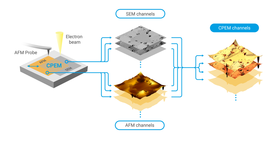
LiteScope AFM-in-SEM offers a wide range of measurement modes to examine different sample properties. Below you can find a list of AFM techniques we offer to complement your SEM measurements, based on our game-changing CPEM (Correlative Probe & Electron Microscopy) technology. CPEM represents a unique value of AFM-in-SEM correlative microscopy, assuring in-situ acquisition & simultaneous seamless correlation of AFM and SEM channels.
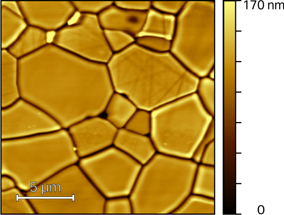
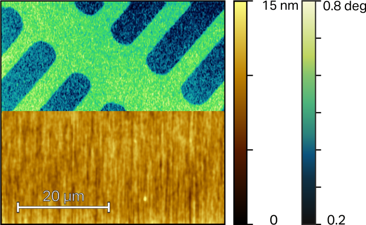
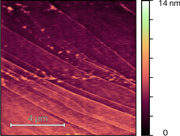
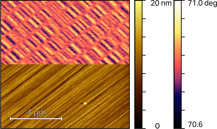
Topography
AFM surface topography allows high-resolution measurements of a wide range of samples. Different types of self-sensing cantilevers can be used. Measurements can be made in contact or tapping mode.
Energy Dissipation
Energy dissipation provides imaging of the local elastic properties of the material. An alternative to FMM, that provides similar information while requiring almost no change from the basic topography measurement setup.
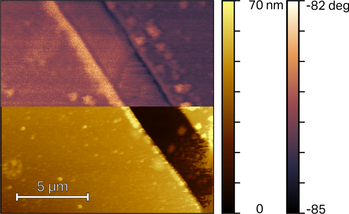
Phase Imaging
Phase imaging is a technique used to map variations in surface properties such as elasticity, adhesion, and friction. It works as one-pass a technique which detects the phase between the probe driving signal and probe oscillation signal while maintaining the constant amplitude of the oscillations.
Nanoindentation
A widely used method for material hardness characterization. LiteScope’s dedicated nanoindentation module from Alemnis enables local hardness and elasticity measurement with supreme control over experiment conditions inside SEM.
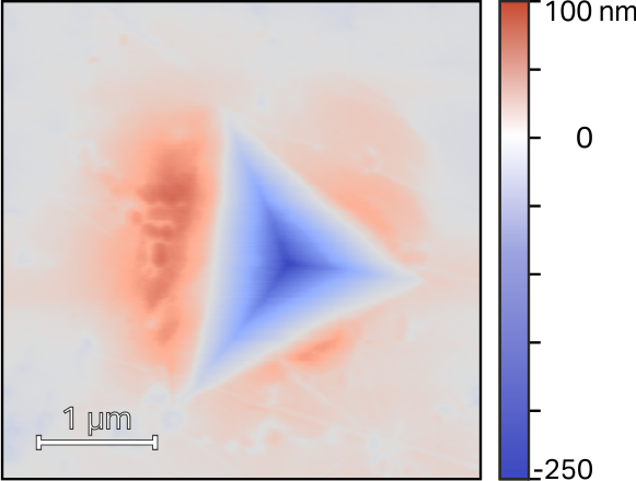
Piezoresponse Force Microscopy (PFM)
PFM allows imaging and manipulation of piezoelectric material domains. This method measures simultaneously topography and mechanical response of the material to the applied alternating voltage. The amplitude and phase of the demodulated signal contain information about the local piezoresponse.
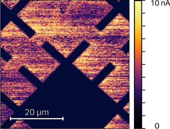
Conductive AFM (C-AFM)
Conductive AFM provides a high-resolution local conductivity map of the sample. A bias voltage is applied between the tip, and the sample and the tip-sample current flow is measured during contact AFM topography measurement.
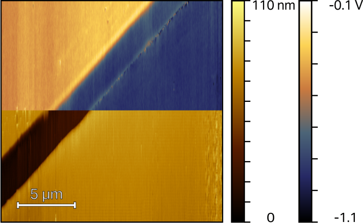
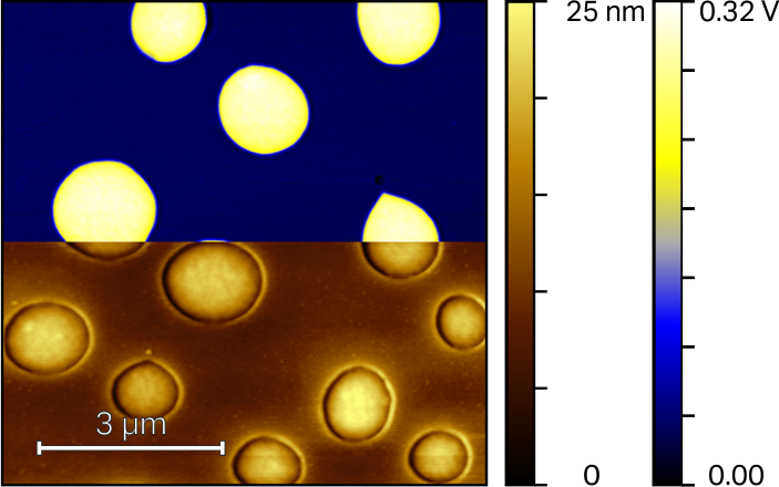
Kelvin Probe Force Microscopy (KPFM)
KPFM is a two-pass technique, estimating the local distribution of surface potentials. In the first pass, the topography in contact mode is measured. The probe is then lifted and repeats its trajectory in the second pass. The electrical interaction between the tip and the sample is then recorded without the influence of topography.
Electrostatic Force Microscopy (EFM)
EFM maps electrical properties of a sample surface by detecting the electrostatic force between the surface and a biased AFM tip which provides useful, qualitative information on electric field gradients of a sample surface.
Scanning Tunneling Microscopy (STM)
STM allows researchers to map the conductivity of a sample‘s surface atom by atom with an ultra-high resolution providing a threedimensional profile of a surface. It enables researchers to examine many characteristics, including roughness and surface defects.
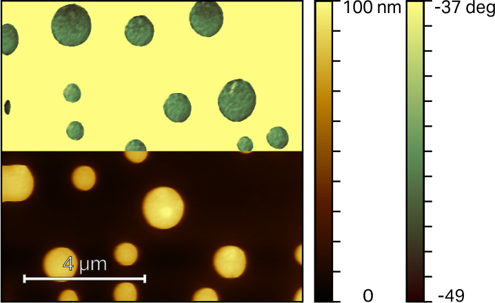
Magnetic Force Microscopy (MFM)
MFM maps the magnetic force gradient above the sample surface. Similarly to KPFM, the topography and magnetic signals are measured in two separate passes over each line.
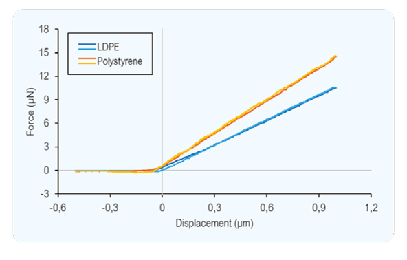
Force-distance curves
F/z spectroscopy is a useful tool for precise local sample characterization. Spectroscopy is used for many purposes like a sample stiffness analysis, detailed surface-tip force progress or local elasticity/plasticity determination.
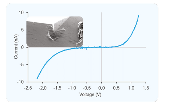
I-V curves
I-V curves give detailed information about the electrical properties of the sample. The AFM-in-SEM configuration provides precise tip navigation and other possibilities for experiment design.
Are you interested? Feel free to...