We are pioneers of in-situ multimodal correlative imaging
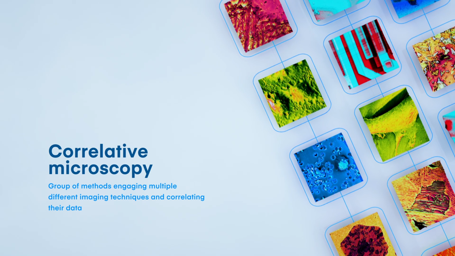
The correlation of images from two microscopes can be limited by the difficult localization of the region of interest or incompatibility of data acquired by different instruments under different conditions. To achieve the best results possible, you need to obtain and variety of AFM modalities (AFM, C-AFM, KPFM, MFM) and SEM images (SE, BSE, EBIC, EDX) at the same time, under the same conditions and in a user-friendly way.
This can be achieved by unique CPEM (Correlative Probe and Electorn Microscopy) technology, developed by NenoVision. It represents a combination of complementary AFM and SEM that enables you to use the advantages of both these techniques via LiteScope AFM-in-SEM.
Want more info? Feel free to...

LiteScope Featured in a Nanoscale Study on Back-Contact-Free Electrical AFM
A new study in Nanoscale introduces electron-beam excited AFM (EB-AFM) — a way to run electrical AFM without a physical back-contact. A low-energy electron beam near the AFM probe acts as a remote electrode, removing the destructive sample preparation that conventional conductive AFM requires.

LiteScope Helps Localize Defects Invisible to SEM in a Joint Study with NVIDIA
A study presented at ISTFA 2025 by NVIDIA and NenoVision introduces a streamlined approach to semiconductor failure analysis. The work integrates in-situ conductive AFM (CAFM) with plasma FIB in a single SEM platform, making it possible to localize electrical defects that conventional SEM imaging can miss.

LiteScope Contributes to Study on Creep Behavior and Microstructure Evolution in Advanced Alloys
A recent study published in the Journal of Materials Science investigates the microstructural evolution and creep behavior of recrystallized FeCr-based alloys. The work combines multiple correlative microscopy techniques to provide a comprehensive view of grain structure, crystallite size, and surface topography.