Correlative atomic force microscopy and scanning electron microscopy of bacteria-diamond-metal nanocomposites
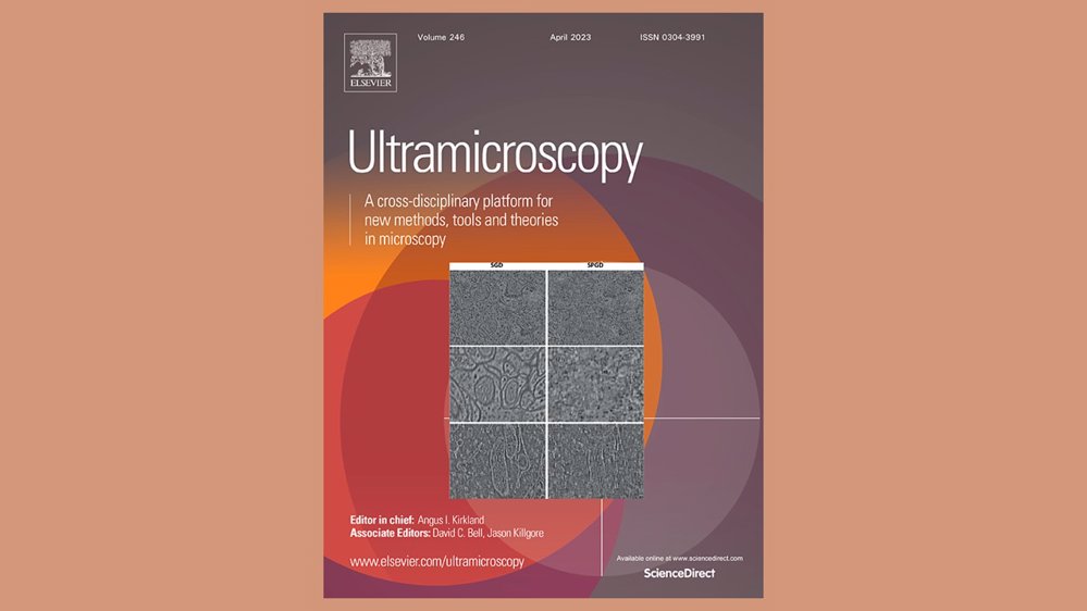
Discover the latest breakthroughs in researching the interface between biological organisms and nanomaterials. This article introduces a groundbreaking approach using AFM-in-SEM LiteScope, a novel combination of microscopic techniques, to unravel interaction mechanisms and effects. Dive into the microscopic realm for a concise exploration of this innovative methodology.
Scientific articles
|
17. 01. 2024
|
by Ultramicroscopy
Product
Technology
Related articles
Scientific articles
|
17. 12. 2024
|
by ACS Applied Electronic Materials
Impact of Electron Irradiation on WS2 Nanotube Devices
Material Science
Technology
Scientific articles
|
18. 11. 2024
|
by Materials
Enhancing Thermoplastic Starch with Maltodextrin: Key Properties and Performance Insights
Material Science
Technology

Scientific articles
|
12. 08. 2024
|
by Scientific Reports
3D Surface Roughness Measurement of Core–Shell Microparticles
Product
Technology

Scientific articles
|
25. 06. 2024
|
by Production Engineering
ZrN coating as a source for the synthesis of a new hybrid ceramic layer
Product
Technology

Webinars
|
12. 03. 2024
Unlocking the Secrets of Battery Materials: A Dive into AFM-in-SEM Characterization Webinar
Material Science
